
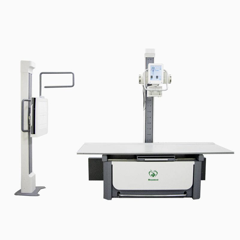
This is the introduction of our whole set X-ray machine,the product type is MY-D023G,here is the specification and characteristic of the product
I. Mechanical Design: Precision and Versatility
The foundation of any advanced imaging system lies in its mechanical architecture. This device adopts a dual-column structural design, which eliminates blind spots during full-body scans and ensures seamless positioning for patients. By integrating the examination bed and X-ray tube column into a single unit, the system significantly reduces installation complexity and minimizes the required (equipment room) footprint. This compact yet robust design is ideal for clinics and hospitals with limited space.
A key innovation is the multi-compatible flat-panel detector tray, which supports detectors from over 80% of manufacturers worldwide. This versatility allows healthcare providers to customize their systems according to clinical needs or budget constraints. Furthermore, the synchronized movement between the detector tray and the X-ray tube column enhances operational efficiency. Physicians can effortlessly adjust imaging angles and positions during examinations, streamlining workflows and improving patient throughput.
II. X-Ray Tube: Power and Reliability
The X-ray tube serves as the heart of the imaging system. This device employs industry-leading X-ray tubes from a manufacturer with 50 years of expertise, guaranteeing unparalleled durability and performance. With a 300 kHU thermal capacity, the tube sustains prolonged operation even under high-demand scenarios, such as emergency departments or busy outpatient clinics.
To achieve diagnostic precision, the system incorporates dual-focus technology (0.6 μm and 1.2 μm focal spots). The ultra-fine 0.6 μm focal spot captures microscopic anatomical details, while the 1.2 μm option balances resolution with broader coverage. Additionally, the adjustable voltage range (40–150 kV) caters to diverse patient demographics and anatomical regions. For example, lower kV settings optimize soft tissue imaging for pediatric patients, while higher kV values penetrate dense structures like adult bone or joints.
III. Flat-Panel Detector: Speed and Sensitivity
The flat-panel detector (FPD) represents a leap forward in imaging speed and clarity. Boasting a data transfer rate of 1,300 Mbps, this detector generates high-resolution images within 3 seconds, minimizing patient wait times and enhancing clinic efficiency. Its high Detective Quantum Efficiency (DQE) ensures superior image quality even at low radiation doses, aligning with modern ALARA (As Low As Reasonably Achievable) principles for patient safety.
A standout feature is the Advanced Exposure Detection (AED) system, which automatically synchronizes with any high-voltage generator. This intelligent function adjusts exposure parameters in real-time, compensating for variations in patient anatomy or positioning. Such adaptability reduces retakes, lowers radiation exposure, and ensures consistent diagnostic outcomes.
IV. Software Suite: Intelligence and Integration
The system’s software platform is a masterpiece of innovation, developed entirely in-house by a unified engineering team. Central to its capabilities is the AED Auto-Exposure Module, which automates calibration and exposure settings with a single click. This feature simplifies operation for technicians while maintaining imaging consistency.
Another breakthrough is the marker-free full-spine image stitching algorithm. Traditional spinal imaging requires physical markers to align multiple X-ray shots, a time-consuming and error-prone process. This system, however, leverages AI-driven algorithms to seamlessly merge images without markers, preserving uniformity in grayscale contrast and anatomical continuity. The result is a comprehensive, distortion-free view of the entire spine, critical for diagnosing scoliosis, fractures, or degenerative conditions.
The software also integrates practical tools such as film printing, diagnostic report generation, and DICOM compatibility, enabling seamless data sharing across hospital networks. By unifying hardware and software development, the engineering team ensures optimal performance, rapid updates, and tailored solutions for evolving clinical needs.
Conclusion: Redefining Diagnostic Excellence
This imaging system embodies the convergence of mechanical ingenuity, component reliability, detector sophistication, and software intelligence. Its dual-column design maximizes space efficiency, while the high-capacity X-ray tube and adaptive detector ensure precision across all patient types. The software’s AI-driven features, from auto-exposure to marker-free stitching, empower clinicians to deliver faster, safer, and more accurate diagnoses.
In an era where healthcare demands are escalating, such innovations are not merely technological triumphs but vital tools for improving global health outcomes. By prioritizing versatility, efficiency, and patient-centric design, this system sets a new standard for medical imaging—one where technology and humanity work hand in hand to illuminate the path to better care.

